Monday, February 29, 2016

Unit 6 Reflection: The Skeletal System

In this unit, we learned about the skeletal system. The essential questions from this unit were:

  • What are the microscopic and macroscopic characteristics of the skeletal system?
  • How do disorders of the skeletal system affect different types or parts of bones?
  • What are the effects of vitamins, cells, and the environment on bone density?
  • How does a bone repair itself after being fractured?
  • What are the defining characteristics of different types of joints?
The macroscopic features of bones include the diaphysis (shaft), which surrounds a hollow medullary cavity, and the proximal and distal epiphyses (flared ends). Compact, or cortical bone, is the smooth, dense bone, while spongy bone is more porous, with cross-hatching supports called trabeculae that help the bone resist stress. There are also two types of bone marrow stored in the bones: red, which produces blood cells and is found in spongy bone, and yellow, which stores energy as fat and is found in the medullary cavity.

At the microscopic level, osteons are what make up bone. They are cylinder-shaped weight-bearing units that run parallel to a bone's axis. They are made of tubes within tubes called lamellae between which are tiny spaces called lacunae, which hold osteocytes (mature bone cells that maintain bone). They also have a central canal that holds nerves and blood vessels to support all of the cells in the bone.
anatomy of a bone (Wikipedia)

Bones are assembled into skeletons, which can be divided into the axial and appendicular skeleton. The axial skeleton is made of the bones along the axis of a body: skull, ribs, sternum, and vertebrae. The appendicular skeleton includes all the rest of the bones, which are attached to the axial skeleton. Bones can also be classified by shape: long, short, flat, and irregular. To help us learn about the skeleton, we did a lab where we dissected an owl pellet, classified the bones we found in it, and tried to assemble them into a skeleton.

Contrary to popular belief, bones do not require just calcium to grow. They also need vitamin K, which can be found in animal-based foods, and vitamin D, which the skin can make when exposed to sunlight. Vitamin D is especially important, since it is needed to absorb calcium so that the calcium can be used to make bones stronger. Severe vitamin D deficiency can lead to rickets, the softening of bones to the point where they may become crooked. Lack of calcium can exacerbate osteoporosis, where the bones become more porous and brittle.

Bone fractures can be classified into many different types. A greenstick, or incomplete, fracture occurs when the bone does not break all the way through. A fissured, or hairline, fracture is a incomplete fracture along the axis of the bone. A comminuted fracture is when the bone breaks into three or more pieces. A transverse fracture crosses a bone perpendicular to its axis, an oblique fracture crosses a bone at an angle, and a spiral fracture occurs when a large twisting force breaks the bone. Simple, or closed, fractures are those that do not puncture the skin, whereas compound, or open, fractures are when the end(s) of the fractured bone(s) break the skin.

After a bone breaks, bone cells called osteoclasts, which build up new bone, and osteoblasts, which break down old bone can repair the bone. Osteoclasts and osteoblasts are also used in bone remodeling, such as when a bone is growing.

 
bone remodeling

Bones attach together at joints, which can be classified by function (syn-, amphi-, and di-arthroses) or by structure (fibrous, cartilagenous, and synovial). Synarthroses are immoveable, amphiarthroses are slightly moveable, and diarthroses are freely moveable. When most people think about joints, like the elbow or the knee, they are thinking about synovial joints, which are diarthrotic. Sutures in the skull are an example of a fibrous synarthrotic joint.

I learned a lot of new things this unit, but there are still a few things I wonder about. For example, if the bone marrow in the medullary cavity is just fat, then is the bone marrow fancy restaurants serve really just fat? What happens to the bone marrow of starving people? Does the body utilize the energy stored in the fat there, and what does this mean for the bones? I would also be interested in learning more in detail about the other types of joints, not just synovial joints.

So far, I have been reaching my New Year's goals of getting eight hours of sleep everyday and not procrastinating on studying for my tests. I hope to continue to keep fulfilling these goals throughout the rest of the year.

Wednesday, February 24, 2016

2016.02.22 Owl Pellet Lab

Today we dissected an owl pellet to study the bones inside. Using a probe and forceps, we broke apart an owl pellet, separated the bones from the fur, categorized them, then tried to identify the organism that was in the pellet.
our owl pellet
After studying the bones in our owl pellet, we concluded that the organism in our pellet was a shrew. We determined by comparing our bones with an identification chart. In particular, we used the skull, mandible, and tibia and fibula to determine that our organism was a shrew. Our organism's skull and mandible resembled the skull and mandible of the shrew in the diagram; furthermore, the skull was 14 mm long and the mandible was 10 mm long, the average lengths of a shrew's skull and mandible. Also, the fused-together tibia and fibula (bottom-right-most bone in the picture on the right below) resembled the tibia and fibula of the shrew in the identification chart.
some bones from the pellet
bones sorted in a chart

Compared to a human skeleton, our shrew skeleton had the same basic parts: skull, vertebral column, ribs, and limbs. These bones all fit in pretty much the same way, and they all perform the same function a human's skeleton does. Some of the bones, like the skull, are made of several fused-together parts, just like a human's.

However, compared to a human skeleton, a shrew skeleton has many differences. For example, the skull and mandible of a shrew are shaped very differently from the skull and mandible of a human. Also, a shrew's pelvis is shaped very differently from a human's (it is longer), since a shrew crawls around on all four feet while a human walks upright. A shrew's tibia and fibula are fused together, where as a human's tibia and fibula are separate bones.


Monday, January 25, 2016

Unit 5 Reflection: The Digestive, Endocrine, and Lymphatic Systems

In this unit, we learned about the digestive, endocrine, and lymphatic systems. The essential questions for this unit were:
  • How are the functions of the digestive system fulfilled by the structures of the alimentary canal and accessory digestive organs?
  • How does the body use certain molecules and biochemical pathways to meet its energy demands?
  • How is diabetes a disruption in fuel metabolism?
  • How do glands use hormones to control various activities of the body?
  • What is the role of the lymphatic structures in the body's defenses and circulation?
The digestive system consists of the alimentary canal (also known as the gastrointestinal track), a long, coiled tube that runs through the body and is open at both ends, and the accessory organs to the alimnetary canal. The organs of the alimentary canal are, in order, the mouth, pharynx, esophagus, stomach, small intestine, large intestine, anal canal, and anus. These organs either digest (break down) food or absorb (take in) nutrients from the food. Assisting them are the liver, gallbladder, and pancreas, which make and store enzyme that help with digestion.
the digestive system (Wikipedia)
The endocrine system is the system of glands in the body that release hormones that affect the body. Hormones can be steroid (lipid-soluble) or non-steroid (non-lipid-soluble) and act like "keys" to "unlock" a certain result or action from a cell. The many glands of the endocrine system include the pituitary, thyroid, parathyroid, and adrenal glands and the pancreas. Each one releases one or more different hormones that regulate different parts of the body.
the endocrine system (Wikipedia)
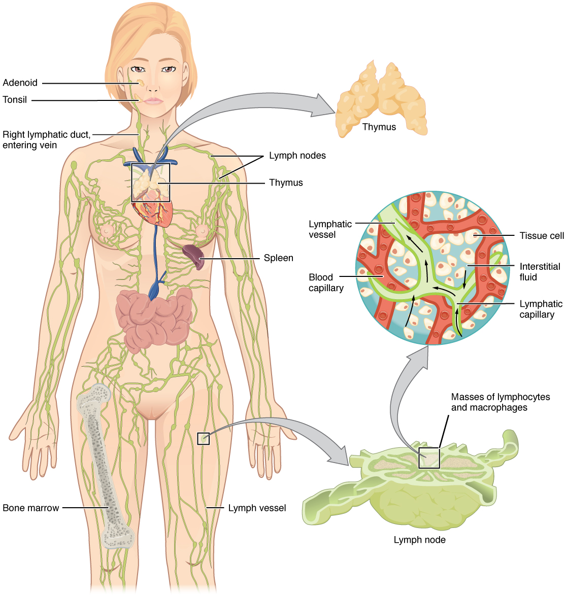
The lymphatic system is a system of vessels that run parallel to the circulatory system's blood vessels and contain a plasma-like fluid call lymph. This lymph comes from liquid lost by the circulatory system or from tissues, and the lymphatic system's job is to return it to the circulatory system. The lymphatic system is also very closely related to the immune system, as there are areas where lymph vessels congregate, called lymph nodes, which host a huge number of immune cells, ready to attack any foreign invader that might be in the lymph. The lymphatic system also includes the thymus, tonsils, and spleen, which are also involved in the immune response.
the lymphatic system (Wikipedia)

One of the themes of this unit is balance. One of the systems we learned about, the endocrine system, is especially all about balance. It makes sure that there is enough of each hormone in the bloodstream at any given time so that homeostasis is properly maintained. The hormones released by the glands of the endocrine system can regulate fluid retention, metabolism, protein synthesis, mineral levels in the skeletal system, heart rate, blood pressure, respiration, and many more. If this delicate hormone balance is upset, it can spell disaster for the body. In this unit we also learned about diabetes, which occurs when the body cannot properly regulate the level of glucose in the bloodstream. This may be because the body cannot produce insulin (type I diabetes) or because the body stops responding to insulin (type II diabetes).

During this unit, we did one lab, the digestive system lab. I thought the lab was very interesting, since it let me see firsthand just how long the digestive system really is. I had always known that the digestive system is very long in the way that one knows a strand of DNA is long, but doesn't actually realize it until one holds it in one's hand.

I would have loved to learn more about the lymphatic system, which most people don't know much about. I am also interested in the different hormones of the endocrine system and how exactly they affect different parts of the body.

At the beginning of this semester I set some New Year's goals, which were to get more sleep and to study for exams earlier. So far, I have gotten more sleep but not as much as I had wanted to (8 hours). However, I am reviewing the material I have learned every day, accomplishing my second goal. I will continue to work on my first goal and hopefully reach it by the deadline I set for myself (February).

Wednesday, January 6, 2016

2016.01.05 Digestive System Lab

Today, we made a measured the length of our digestive system. The main organs of the digestive system are the mouth, the esophagus, the stomach, and the small and large intestines. We used different colors of ribbon to represent each organ in the digestive system. I chose blue ribbon to represent the mouth, green to represent the esophagus, pink to represent the stomach, white string to represent the small intestine, and red to represent the large intestine. Then I tied all the pieces together, resulting in a very long string, representing the length of my digestive system.
lengths and color of strings I used
all the string I used
The length of my digestive system turned out to be 8.336 meters. In comparison, my height is 1.524 meters, which means that my digestive system is almost 6 times my height. In order to fit such a long organ system in my body, it must be folded over and over again (especially the small intestine, which is the longest organ in the digestive system).

I though that it takes about 2 days for food to move through the entire digestive system. After searching online, I found that it takes a little less than 2 days (30-40 hours) for food to move through the entire digestive system, which is pretty close. Factors such as the composition of the food, gender, and stress levels all can affect the amount of time it takes for food to move through the digestive tract.

Digestion is the breaking down of food into little pieces (nutrients), while absorption is the taking in of these nutrients by the body. Digestion occurs in the mouth and stomach, while it is the intestines that absorb nutrients from the digested food.

Monday, January 4, 2016

New Year's Goals

I have set some goals for 2016:

  • I will get at least 8 hours of sleep everyday. To do this, I will manage my time better, such as by allocating a specific amount of time for everything I do, so that I do not waste any time and have more time to sleep. I will achieve this goal within by February.
  • I will start studying for tests earlier. After every class, I will review what I have just learned, instead of just studying for tests the night before. This will allow me to retain information better, as well as give me less exam-related stress. Next time I have an exam, I won't be cramming, and I won't be losing any sleep by staying up later to study for a test.

Monday, November 16, 2015

Sheep Heart Dissection Video Tutorial

After we finished our sheep heart dissection, we made a video explaining the parts of the heart, their functions, how the blood flows through the heart, and how the heart beats. The video is below and can also be found on YouTube here and here:

Thursday, November 12, 2015

2015.11.01 Sheep Heart Dissection

Today we dissected a sheep's heart. We were able to observe all the structures on the outside and inside the heart. As we studied the heart we answered questions that our teacher gave to us. The answers are below, and the question numbers are in parentheses ().

On the outside of the heart is the pericardium, a two-layered membrane sac that protects the heart and secretes a fluid that lubricates the heart to reduce friction. It also keeps the heart together as it continuously contracts and relaxes (1).

There are many blood vessels that connect to the heart. Blood vessels that take blood from the heart to the body are called arteries. Blood vessels that take blood from the body to the heart are called veins. Arteries are more elastic than veins, and their walls are thicker and contain more smooth muscle than the walls of veins. This lets the arteries expand and contract under the pressure that the heart exerts against the artery walls as it pumps blood (2).

The inside of the heart is divided into four chambers, two atria and two ventricles. The four chambers are divided into left and right. Each side (left or right) has one atrium and one ventricle. The right side of the heart pumps blood only to the lungs, so it is not as large and muscular as the left side of the heart, which pumps blood to the whole body. The right side of the heart contains deoxygenated blood, while the left side of the heart contains oxygenated blood (12).

The atria are the superior chambers that receives blood from the veins and pumps it into the ventricles. On top of each atrium is a dogear-like pouch that increases the blood volume of the atria so they can receive more blood from the veins (3). Since the atria only have to pump blood to the ventricles, their walls are not as thick and muscular as the walls of the ventricles, which have to pump blood to the lungs or to the whole body (4).

The atria are separated from the ventricles by atrioventricular valves, which prevent blood in the ventricles from flowing back into the atria when the ventricles contract. The atrioventricular valves, which are the tricuspid and bicuspid (mitral) valves, are "anchored" by the chordae tendinae, which are long fibers that trail off the valve, and the papillary muscle, which attach the chordae tendinae to the wall of the ventricle. This makes sure the valves close properly and keeps the valves from swinging backwards into the atria so that blood does not leak backwards into the atria. If valves are not anchored properly, blood could leak backwards instead of going where it needs to go, which could cause health problems like fatigue, swelling, or heart palpitations (irregular heartbeat) (7).
drawing of the tricuspid valve,
showing the chordae tendinae and papillary muscle (6)

drawing of the bicuspid (mitral) valve,
showing the chordae tendinae and papillary muscle (8)
The semilunar valves are the valves that separate the ventricles from the arteries that leave the heart. Just like the atrioventricular valves, they prevent blood from flowing backwards. The semilunar valves prevent blood flow back into the ventricles and keeps the blood flowing in the arteries away from the heart (9).
drawing of aortic semilunar valve (11)

Valve failure on different sides of the heart has different effects. If valvular heart disease occurs on the right side of the heart, swelling in the feet and ankles occurs because blood that is pumped into the right ventricle by the right atrium does not stay in the right ventricle. Instead, it flows back into the right atrium, backing up the blood flow and not letting blood from the body flow into the right atrium. As a result, blood collects in the feet and ankles, causing them to swell up (10a).

If valvular heart disease occurs on the left side of the heart, the body would not be able to get as much blood. Body cells would not get as much oxygen, which would cause fatigue. This is because blood in the aorta that is supposed to be pumped to the body by the left ventricle instead flows back into the left ventricle and does not go to the body (10b).

At the very end of the dissection, we made a frontal cut through the heart to separate the anterior (front) and posterior (back) sides. This way we were able to see all the structures inside the heart.
drawing of interior of heart (13)

Tuesday, November 10, 2015

Unit 3 Reflection: The Circulatory System

Unit 3 was about the circulatory system and the respiratory system. The essential questions of this unit included:
  • What are the major features of the circulatory system?
  • What are the structures of the heart and their functions?
  • What are the (dys)functions of the blood vessels?
  • What are the different components of blood and what are their functions?
  • What are heart attacks and strokes? What causes them, how are they identified, and how are they treated?
  • What are the functions of the respiratory system?
The circulatory system is a fluid-filled network of tubes throughout the body. It is made of several major components: the heart, which pumps the fluid through the system; the blood vessels, which carry the fluid through the system; and the blood, which is the actual fluid. There are also several accessory organs to the circulatory system: the lungs, which exchange carbon dioxide in the blood for oxygen; the kidneys, which remove excess waste and water from the blood; and the intestines, which absorb nutrients and water from digested food into the blood.

We learned about different parts of the heart in detail in this unit. To help us learn about the heart's structure, we went outside, drew a diagram of the heart on the ground, and made a video explaining the path a red blood cell takes through the heart.
Our diagram (top) and video (left) of the heart
I also drew a diagram of the heart by myself to help me learn about the heart. This was after we had learned more about the structures of the heart and their functions, so it is more accurate (the papillary muscle in the above diagram is incorrectly labeled).
my diagram of the heart
All this drawing of the heart helped us prepare for our sheep's heart dissection, which was very interesting and let us see what an actual heart looked like. There was some fat on the heart, which we did not really expect because diagrams of the heart don't really depict fat, and the anatomy of the heart was a little bit different since it wasn't a human heart.
anterior (front) view of sheep's heart
(where's the inferior vena cava!?)

heart cut lengthwise
(the way most heart diagrams are drawn)
We also learned about blood vessels. There are three main kinds of blood vessels: arteries, which take blood from the heart to the body; veins which take blood from the body to the heart; and capillaries, which connect the two. Arteries are the ones that expand and contract as the heart beats (this is how you feel you pulse). Veins have valves in them that prevent blood from flowing backwards (e.g. blood in your leg has to flow against gravity to get back to your heart). The capillaries are where the exchange of nutrients, wastes, and gasses actually occur.

In the blood vessels flow the blood, which is made of plasma and cells. Plasma itself is made of water and dissolved solutes. Blood cells include red blood cells, which carry oxygen, and white blood cells, which are part of our immune system and help fight infections. There are many different kinds of white blood cells, including neutrophils, basophils, eosinophils, lymphocytes (B-cells and T-cells), and monocytes (which mature into macrophages). Blood also contains platelets, which help the blood clot if you get cut.

What we learned about heart diseases tied into our past unit about health. Mostly we learned about atherosclerosis (the buildup of plaque, or fatty material, in the blood vessels) and aneurysms (the weakening of a blood vessel wall due to blood getting in between the layers of the wall and making it swell up). Atherosclerosis is caused by chronic inflammation; excess sugars, omega-6, and LDL ("bad") cholesterol; and too little HDL ("good") cholesterol. When little microtears form in the blood vessel wall, this excess LDL becomes stuck in the wall. If there is too little HDL to clean it up, white blood cells think that the body is under attack, come to engulf the LDL, and become stuck as well. This forms a plaque in the artery that restricts blood flow. A blood clot can form around the plaque and cut off blood flow entirely. If this happens in a coronary artery that provides blood to the heart muscle, it is called a heart attack. If this happens in the brain, it is called a stroke (strokes can also be caused by aneurysms). Good nutrition lowers your risk of atherosclerosis (since atherosclerosis is caused by excess sugars, omega-6, and LDL) and therefore the diseases associated with it. Exercising, managing stress, and controlling your weight also prevents atherosclerosis.

We also learned about the respiratory system, since it is connected to the circulatory system (oxygen-poor blood is pumped from the heart to the lungs to exchange carbon dioxide for oxygen, then returns back to the heart to be pumped to the rest of the body). Air enters the respiratory system through the nose, then travels down the pharynx (throat), larynx (voice box), trachea (windpipe), bronchus, broncial branches, and finally into the lungs. The lungs themselves are made of tiny air sacs called alveoli, which are covered with capillaries and are where the actually gas exchange takes place. Breathing in occurs when the diaphragm contracts and flattens out from its relaxed, curving-upward state (this is interesting; I didn't know about how the diaphragm actually moved down before), and the intercostals (muscles between the ribs) contract and pull the ribcage out.

This unit was a pretty good unit for me, since I understood what was taught in class. I felt more comfortable with this unit's material than with last unit's on health; I suppose I am better at learning about the larger things that I can see and feel than I am at learning about the little things that I can't really see for myself. The active studying method that we discussed in class also helped, especially relating concepts to each other by drawing concept maps.
active studying
 Last unit we made a chart of health goals. My goals were to:
  • eat seafood at least three times a week.
  • exercise at least three times a week.
  • sleep at least eight hours every day.
So far, I have achieved the eating and the exercise goals. These were pretty easy since my whole family likes to eat seafood, and whenever I wanted to exercise I would just go run with my friends on the cross-country team. However, now that the cross-country season is over, I need to find time during the weekends to exercise with my family or convince some friends to run with me after school (most of them don't really want to run after the season is over), since I don't like to exercise alone. I also have to work harder on getting eight hours of sleep daily. I get eight hours of sleep during the weekends now, but sometimes during the weekdays I stay up later doing homework. I can manage my time better and spend less time lying around so that I can finish my homework earlier and go to sleep earlier.

Wednesday, October 14, 2015

Monday Wellness Reflection: Tea

Our Monday Wellness was on tea. The presentation can be found here.

We chose to do my Monday Wellness project on tea because we had heard that teas had a lot of health benefits. However, I did not know why exactly teas had such health benefits. Also, I knew that there are thousands of different blends of tea and had heard that different teas have different health benefits. We had also heard of how antioxidants in tea were good for the body, but we did not know what antioxidants actually were; we also heard that tea was a good substitute for coffee, but beyond "because it is more healthy," we did not know exactly why.

When researching for my Monday Wellness, I found it interesting that all "true" teas (white, green, oolong, black, pu'er) are made from the same plant, Camellia sinesis. I had previously thought that different teas came from different plants (e.g. green tea came from green tea plants, black tea came from black tea plants, etc.). The different teas and their distinct characteristics come from the different ways the plant is processed.

We also finally learned how exactly antioxidants and caffeine affect our bodies. We always hear that antioxidants are good and that caffeine is bad, but many people do not understand why this is so. Antioxidants actually work by keeping harmful particles in our body, called "free radicals," from being harmful. Caffeine is actually a naturally occurring chemical compound in some plants that is a stimulant drug in humans. It was very interesting to find out that caffeine in moderation is actually good for the body, since it increases mental alertness, improves memory, and reduces the risk of certain diseases.

The most important theme of our Monday Wellness was moderation. We also talked about moderation and balance in class during our health unit. Sure, drinking tea is good, but drinking too much for too long may result in side effects. In fact, drinking too much of anything, even water, which is essential for life, is harmful. Furthermore, "too much" is different for everyone depending on their age, gender, body weight, and many, many other factors; it is not a concrete number. We frequently want to hear numbers so that we know what we should or should not do -- I saw this when people started asking me about how many cups of tea they should drink and how many cups was too much -- but we should keep in mind that there are not really any clearly set limits that fit everyone.

On a scale of 1 to 10, I would give us an 8. Since we did not practice our presentation together before actually presenting it in class, our timing was a little off. It took more time than we expected to set up and clean up our activity, so in the end I did most of the presentation while my partner set up and cleaned up the activity, since she brought the supplies for it. We also wanted to do the activity before most of the presentation to wake up the students and get them more involved in the presentation. However (as stated before), it took longer than we expected to set up the activity, so we got through half of our presentation first before we did the activity. As expected, the activity did wake up the students and get them more involved: during the half of the presentation before the activity, I felt like the students were just staring at me talk and not engaging; after the activity, the students seemed much more lively and asked a lot more questions.

Tuesday, October 13, 2015

2015.10.13 Measuring Blood Pressure and Pulse

Today we learned how to take blood pressure and pulse and practiced doing so on each other.

Blood pressure is the pressure blood exerts on the walls of arteries when the ventricles of the heart contract and send blood into the body. There are two parts of blood pressure, systolic pressure and diastolic pressure. Systolic pressure is the pressure that the blood exerts on the walls of the arteries when the ventricles contract, so it is higher. Diastolic pressure is the pressure that the blood exerts on the walls of the arteries when the ventricles relax, so it is lower.

Blood pressure can be measured using a digital sphygmomanometer (blood pressure cuff), or using a manual one and a stethoscope. In class we learned how to use a manual sphygmomanometer and a stethoscope to measure blood pressure:
  1. Put the patient's arm palm up on a flat surface. Make sure the patient's arm is level with his heart.
  2. Wrap the blood pressure cuff around the patient's upper arm, at least 1 inch above his elbow.  The tubes that connect the pressure gauge and the pump to the cuff should be pointing down, in line with the patient's arm. Secure the cuff.
  3. Put on the stethoscope. Put the flat part of the stethoscope on top of the brachial artery, on the inside of the patient's arm a little above his elbow. You will not be able to hear anything yet.
  4. Make sure the valve on the pump is closed. Start pumping air into the cuff until the pressure gauge reaches about 150 mmHg.
  5. Slowly release air from the cuff by opening the valve on the pump slowly. The needle on the gauge should fall about 2 mmHg every time you open the valve.
  6. Note the number on the gauge when you hear a very faint tapping sound through the stethoscope. This is the systolic pressure.
  7. Keep releasing air from the cuff until you stop hearing anything through the stethoscope. The number on the gauge at which this occurs is the diastolic pressure.
  8. Blood pressure is written as a fraction, systolic pressure/diastolic pressure.
We tried taking blood pressure using a sphygmomanometer and a stethoscope on each other:

Blood Pressure Results


Subject 1
Subject 2
Trial 1
90/50
110/80
Trial 2
100/60
105/70

Pulse is the number of times the heart beats over a certain amount of time. It can be measured by using a stethoscope over the heart, or just with 2 fingers (index and middle) over the radial artery (in the wrist) or over the common carotid artery (in the neck). Count the number of times the heart beats for 15 seconds, then multiply that number by 4. We do not use the thumb to measure pulse because there is also a pulse in thumb, which may throw off how many times we count the heartbeat. We tried taking pulses at different locations on each other:

Pulse Results

Subject 1
Subject 2
Average for All Subjects
Radial
70
70
70
Carotid
68
70
69
Heart (stethoscope)
72
86
79
Average of Individual Subjects
70
76
73