In class we learned about four main types of tissue: muscle tissue, which allows for movement; epithelial tissue, which covers our body and lines our hollow organs; nervous tissue; and connective tissue, which connects all other kinds of tissue. All these different kinds of tissues can be separated into even smaller categories. For example, muscle tissue can be separated into smooth, skeletal, or cardiac. Epithelial tissue is classified by its cells' shapes (squamous, cuboidal, columnar) and layering (smooth, stratified, pseudostratified). Nervous tissue includes both neurons and neuroglial cells that support the neurons. Connective tissue in particular is an extremely broad category, ranging from blood to bone to the extracellular matrix around cells.
Today we studied different kinds of tissues under the microscope. It was very interesting to see what different tissues looked like in real life (and sometimes kind of frustrating, because in real life, tissues do not fall neatly into one category or another). For the most part, however, the nuclei in all the tissue samples were easily seen, which helped a lot with identifying the tissues. It also reminded me that the tissues we learned about in class are not all the tissues in the body, which is easy to forget because -- well, that's all we learned about.
Of course, there were some tissues that were taught in class and looked exactly the same in diagrams as under the microscope. For example, identifying muscle tissue was pretty easy:
 |
| smooth muscle cells |
|
 |
| skeletal muscle cells |
|
 |
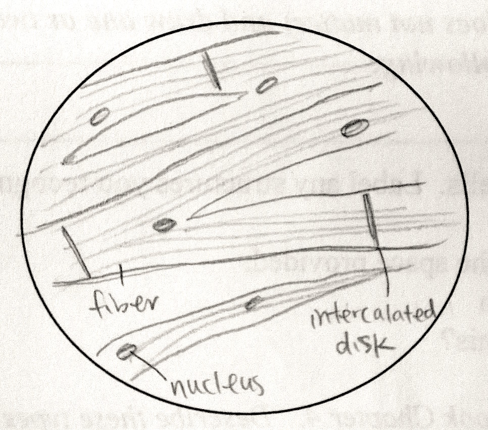
| cardiac muscle cells |
|
The unique spindle-shaped cells in the smooth muscle were very easy
to see, and the striations (protein fibers) in skeletal and cardiac
muscle that distinguish them from all other tissues were readily
apparent. Furthermore, the branching structure in cardiac muscle and the
intercalated disks that keep the cardiac muscle cells together under
the strain of making the heart constantly were very obvious.
However, take this example:
 |
| like, what in the world is this supposed to be? |
This is from the skin of a human. The top layers (1) are stratified squamous epithelial tissue, which we learned in class makes up our skin. The bottom layers (2) are connective tissue, which connects other kinds of tissues together (in this case, the skin to the rest of the body). Since we don't want our skin to be falling or sagging off our body, this particular kind of connective tissue is dense irregular connective tissue.
One of the (I think) most interesting ones was the tissue from the aorta. It is also made of connective tissue, but this kind was elastic connective tissue, which means that there are more elastic fibers than collagenous fibers. This makes sense because the aorta is where blood is pumped out of the heart and must be able to stretch to accommodate the pressure of the blood being pumped through it.
And just thought I'd put my favorite of all the tissue samples (it looks a lot more impressive in real life):
 |
| compact bone |


No comments:
Post a Comment